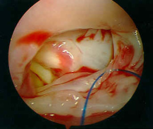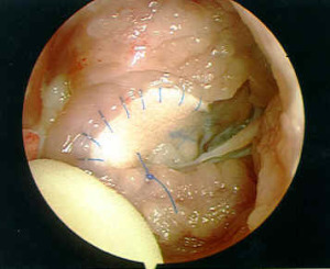

Tetralogy of Fallot is caused by an abnormal location of the muscle that separates the aortic valve and the pulmonary valve in the normal heart (anterior and cephalad displacement – malalignment – of the infundibular septum). This has several effects: (Figure)
- obstruction of pulmonary blood flow
- creation of a ventricular septal defect (malalignment VSD)
- consequent hypertrophy of the right ventricle, caused by obstruction to pulmonary blood flow
- underdevelopment of the pulmonary arteries and small pulmonary arteries
Tetralogy of Fallot is a spectrum of anatomy, with varying degrees of pulmonary obstruction, and different levels of ventricular hypertrophy.
In addition, Tetralogy of Fallot may be associated with other conditions: atrial septal defect, patent ductus arterious, atrioventricular canal defect, and muscular ventricular septal defects
Physiology
The obstruction to pulmonary blood flow may be intermittent because the obstruction may be partially dynamic (changeable depending on the particular hemodynamic state). Reduced pulmonary blood flow causes decreased oxygenation of the blood (cyanosis or “blueness”). Infants with Tetralogy of Fallot rarely have episodes of cyanosis (“Tet spells”). Cyanosis typically increases in frequency with age as the right ventricular becomes increasingly hypertrophied.
Pathology
Decreased oxygenation can have deleterious effects on the function and development of vital organ systems such as the brain and the heart itself, although somewhat low oxygen levels may be tolerated for some time. A severe “Tet spell” may reduce oxygenation to such a low level that the baby may not survive the episode. Tetralogy tends to worsen over time, as pulmonary outflow obstruction causes worsening right ventricular hypertrophy and the subsequent increase right ventricular hypertrophy causes more obstruction to pulmonary blood flow. Right ventricular hypertension and hypertrophy may eventually lead to RV failure and severe arrhythmias.
Surgical Indications and Approach
Tetralogy of Fallot requires surgical repair. Repair is accomplished, in the usual circumstance, electively between two and six months of age. Repair is indicated at any time when the child is symptomatic or has “Tet spells”. Repair requires:
- closure of the ventricular septal defect with a patch
- relief of pulmonary outflow tract obstruction, which may8 require resection of right ventricular muscle bundles, pulmonary valvotomy or excision
- repair of associated defects